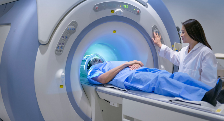
If you are prescribed tomography of the abdominal cavity, it is important to conduct the correct preparation. Read more in the article.
Contents
- What is a computed tomography method, MRI?
- Indications for examination using the tomography of the abdominal cavity
- What is important preparation for CT (tomography) of the abdominal cavity?
- What is allowed to eat and drink before tomography of the abdominal cavity?
- Features of preparation for tomography of the abdominal cavity using contrast: What is the meaning of the method?
- The negative effects of the tomography of the abdominal cavity on a person: contraindications of the method with contrast and without it
- Video: Preparation for MRI: How to prepare for an MRI of the abdominal region and a small pelvis?
Almost every person is prescribed to go through an MRI of different organs. This is one of the most popular types of examination. Often doctors prescribe tomography when you need to learn about the state of health of the abdominal cavity. This is the most informative method. What is this procedure? Why do you need to undergo mandatory preparation for MR tomography of the abdominal cavity? How to be prepared? Will this procedure harm the health of the patient? Look for answers to these and other questions in this article.
What is a computed tomography method, MRI?
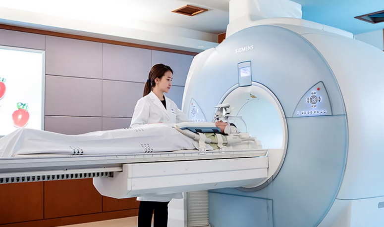
Magnetic resonance imaging is the most popular type of examination. This painless computer method is characterized by informativeness and reliability. MRI makes it possible to detect even minor changes or deviations from the norm in the body that could not be identified using other types of examination.
A similar level of accuracy is provided by the scanning of soft or hard tissues, blood vessels, the layers of which are applied to each other, displaying the real picture of the state of organs. Doctors have the opportunity to explore the images of the abdominal and retroperitoneal space from different positions.
Indications for examination using the tomography of the abdominal cavity
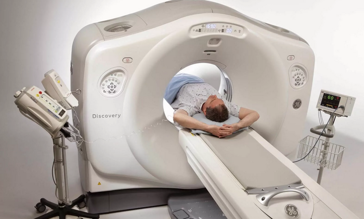
This technique allows you to explore all organs and systems of the abdominal cavity:
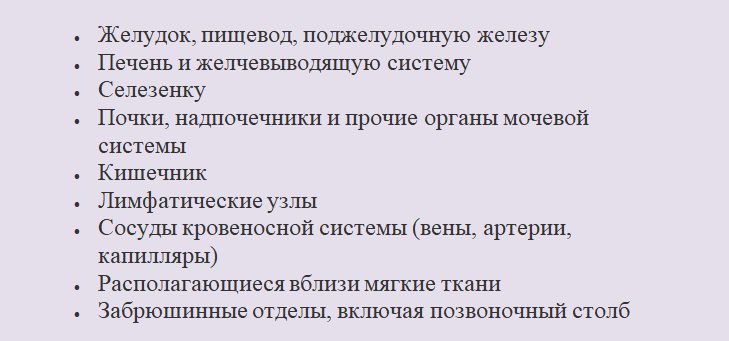
Indications for research With the help of thomography of the abdominal cavity are different:
- If other types of examination (ultrasound, radiography) are unsuccessful
- Assumptions that there are malignant tumors and metastases on the surface and inside organs and the spinal column
- Acute or chronic inflammation, fluid accumulation
- Expanded spleen or kidneys
- Damage that entailed injuries of organs and caused internal bleeding
- Deviations in the structure or development of the structures of the body
- Benign cysts, adenoma
- Deviations from the norm
- Deterioration of blood circulation (ischemia)
- Kidneys and gall bladder stones
- Inflammation of the pancreas
- After surgery, in order to assess the patient's condition or in case of complications
- If this is the only possible type of research
- Abdomen
- Anomalous structure of internal organs
- Symptoms of unidentified etiology
- Surgery planning
- Assessment of the effectiveness of selected treatment
Scan allows you to perform diagnostics:
- Injury of internal organs, vascular mesh, tissues
- Inherited or received deviations
- Arrangement of organs
- Benign and cancer tumors, metastases
- Inflammation, obstruction or degenerative processes
- The effectiveness of the performed surgical intervention or the presence of complications (adhesions, scars, tissue inflammation)
- Circulatory disorders
- Deviations from the norm of blood vessels
- Violations in nerve roots, trunks
- The presence of deposits in gallstone and urinary systems (gall bladder, kidneys)
- Violations of the structure of the spine in the lower back and middle part of the back
The content, the provision of accurate data and the safety of the examination make it more and more in demand. Often, MR is performed for preventive purposes.
What is important preparation for CT (tomography) of the abdominal cavity?

The abdominal department is a large part of the human body in which different inflammatory or destructive processes can occur. In view of this, there are a certain number of MRI categories that make it possible to examine how it proceeds and at what stage a specific disease is located.
- Viewing CT tomography is the most complete. It allows you to examine the part of the body in the belly area as a whole (the location and condition of organs, blood vessels, soft tissues).
- In case of violation of the blood flow, angiography or CT of the venous sinus is used.
- The examination can be carried out with the use of contrast or without it.
What is important preparation for tomography of the abdominal cavity? Any type of examination requires correct preparatory measures. This will contribute to obtaining the most accurate and correct results, taking into account the operation of the device and the functionality of the diagnosed part (it is important that all intestinal parts are as empty as possible). Preparatory measures for MR tomography are quite simple. The patient needs to complete the simple instructions of the doctor for several days:
- At home, it is worth observing a simple diet, its essence will be described below.
- The preparation itself will not require additional financial and physical costs.
- Before the CT process, the patient puts on a disposable robe or medical shirt. In most clinics, it is provided free of charge, in some cases it is separately bought by a patient.
- The most important thing is to remove all metal items - jewelry, piercing, clocks, hairpins, dentures, etc. Any metal object will be attracted by a large magnetic element, the result of which can be a breakdown of expensive equipment.
It is worth knowing: The procedure is unacceptable for people with non -removable metal plates containing metal, as well as pacemakers and insulin pumps.
What is allowed to eat and drink before tomography of the abdominal cavity?

During the initial consultation, the doctor explains the importance of dietary education and develops the diet. It is important to follow it within 2-3 days. Such nutritional features before the abdominal cavity are important to avoid the formation of gases and for better liberation of the intestinal departments. The food should be based on natural and light foods with a slight carbohydrate content.
Which should not be consumed:
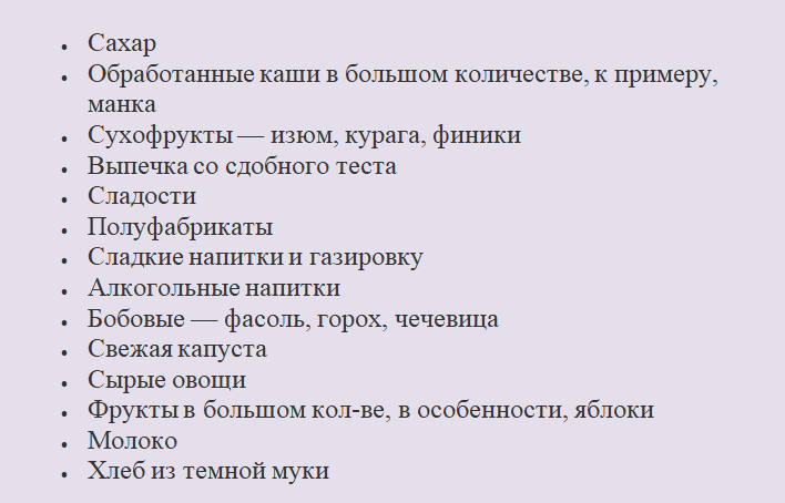
To still reduce gas formation, it is advisable to use specially designed drugs for this:
- Espumisan
- Potzaz
- Smecta
- Neo -Mectin, etc.
Food should consist mainly of these products:
- Stewed, boiled or baked vegetables in the oven
- Low -fat fish
- Meat or seafood
- Greenery
- A small number of berries
- Boiled eggs
A moderate amount of fermented milk products, for example, cottage cheese with a small amount of fat components, classic ugly yogurt is allowed. You can eat porridge with a slight content of a number of carbohydrates- buckwheat, brown rice, oatmeal cereal (not flakes, but whole grains), yachka and wheat cereal. As drinks - water or tea without sugar.
The menu is developed by the doctor separately for each patient. The state of health and other characteristics of the human body affect nutrition. For 6-8 hours. Before the research process, it is not allowed to eat. The use of fluid is also limited. In 4 hours. Before the process, you can drink 1 tbsp. water, but no later.
Features of preparation for tomography of the abdominal cavity using contrast: What is the meaning of the method?
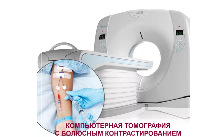
An examination can be carried out using a contrasting substance or without it. As a rule, an examination is carried out without this drug, since such a method already provides all the data necessary for diagnosis. Contrast is used for suspected different formations of a benign or malignant nature, different pathologies, inflammation. The meaning of the technique is simple:
- A special drug (contrast) is injured inside the vein. It instantly spreads with blood circulation.
- During scanning on CT, the product in the vascular grid glows, which is noticeable on the screen.
- Education and foci of inflammation are covered by a huge number of vessels, more than healthy fabrics. In other words, on the monitor they will be brightly highlighted.
- With the help of this method, doctors can determine the nature of a new education in the body (benign or malignant), as well as to say about its exact parameters and location, internal structure and outline, the presence of metastasized elements.
- This makes it possible to detect oncological pathology in the very initial stages, which increases the chances of successful treatment.
Preparation for this examination using contrast is somewhat different from the standard CT of the abdominal department. Here are its features:
- Doctors are required to provide for the presence of a reaction to hypersensitivity to this drug and, if necessary, change it to another tool.
- The patient needs to talk about his allergies to drugs, transfer the cuts of the required tests, and report on previous diseases or operations.
- The doctor should know which tablets are currently accepted, as it happens that the contrast is mutually exclusive to the use of other medicines.
- Be sure to follow a diet. The diet should consist of useful natural products with a small content of carbohydrates that do not provoke gases.
- Patients who have a tendency to disorders of the digestive system and chronic constipation need to make an enema for cleaning. When conducting an MRI, the intestines must be empty. In this case, it will be possible to get the most accurate and untreated information.
- Before the examination, it is advisable to take antispasmodic drugs - No-shpa, spasmalgon.
Patients with psycho. Disorders, constant strong excitability or phobia of a closed space, is shown taking sedative and relaxing drugs.
The negative effects of the tomography of the abdominal cavity on a person: contraindications of the method with contrast and without it
MRI - One of the safest examination methods. The impact of radiation ions is completely excluded, in other words, the patient is not exposed to irradiation. The effect of the magnetic element does not affect the body and the processes occurring in it. With the correct conduct of this method of research in compliance with all norms and rules, it does not harm the human body (neither now nor later).
It is worth knowing: The negative effect of the tomography of the abdominal cavity on a person is completely absent.
Sometimes undesirable reactions to the contrasting drug happen, but this rarely happens. Be sure to inform the doctor about his health and the presence of allergies. As such a contrast is safe. It does not affect the functioning of organs, is not absorbed into the blood circulation and tissue, is quickly excreted from the body. It implies individual immunity of the drug. In such a situation, it should be replaced.
To avoid adverse reactions and deterioration of health, it is necessary to pay attention to the list of states in which the procedure is unacceptable. Here are contraindications to the MRI method with and without contrast:
- The period of pregnancy-the 1st trimester is considered the most dangerous, diagnostics are carried out with emergency.
- The breastfeeding period for the contrast method - the drug may be in milk and harm the child.
- Heart and renal failure.
- Mental illness, since soothing medicines are administered before the examination.
- Implanted prostheses, plates, metal implants, as well as braces.
- The implanted pacemaker or insulin pump - the electronic device will immediately break and there is a risk of injury to the tomograph.
- Large growth and weight of the patient, exceeding 150 kg.
To prevent the examination, and its results were more informative and accurate, you should clearly adhere to the recommendations and appointments of the doctor. The correct preparation for the procedure and immobility of the patient during scanning - within an hour is of great importance, depends on the type of examination. Good luck!

